
Grants & Private Funding
Ongoing Major Private Research Support
Mary Rodes Gibson Foundation for Research in Hemostasis and Thrombosis (2001-present)
Mabel and Everett Hinkson Memorial Fund for Bleeding and Clotting Research (2010-present)
- Molecular linkages between von Willebrand factor (VWF) and the alternative complement pathway (AP) were recently discovered by Nancy Turner and Joel Moake (PLOS ONE, 2013: e59372). Endothelial cell (EC)-anchored ultra-large (UL) VWF multimeric strings function as an activating surface for the AP. C3 (in active C3b form) binds to the EC-anchored ULVWF strings, and promotes the assembly of C3bBb (C3 convertase) and (C3b)2Bb (C5 convertase). These linkages help to explain enigmatic clinical problems related to thrombotic microangiopathies, including: some cases of refractory thrombotic thrombocytopenic purpura (TTP); TTP associated with only mild-modest deficiencies of ADAMTS-13; the provocation (or exacerbation) of acute episodes in patients with the atypical hemolytic-uremic syndrome (aHUS); and thrombosis in paroxysmal nocturnal hemoglobinuria (PNH).
- Recent experiments by Leticia Noalasco and Joel Moake have also demonstrated that complement factor H (FH) performs a dual role: participating in regulation of the AP by binding to EC-anchored ULVWF strings; and functioning as a reductase to decrease the size of soluble VWF multimers (Arteriosclerosis Thrombosis Vascular Biology, 2013; 33: 2524-8).
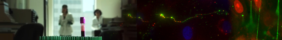
- The cellular synthesis site and ensuing storage location for human factor VIII (FVIII), the coagulation protein deficient in hemophilia A, has been elusive. FVIII stability and half-life is dependent on non-covalent complex formation with VWF to avoid proteolysis and clearance. VWF is synthesized in megakaryocytes and endothelial cells, and is stored and secreted from platelet alpha granules and Weibel-Palade bodies of endothelial cells. Nancy turner and Joel Moake recently provided direct evidence for FVIII synthesis in 2 types of primary human endothelial cells: glomerular microvascular endothelial cells (GMVECs) and umbilical vein endothelial cells (HUVECs) (PLOS ONE 2015: e0140740),/a>. Gene expression quantified by real time PCR revealed that levels of F8 and VWF are similar in GMVECs and HUVECs. Previous clinical studies have shown that stimulation of vasopressin V2 receptors causes parallel secretion of both proteins. In this study, Turner found that both endothelial cell types express AVPR2 (vasopressin V2 receptor gene) and that AVPR2 mRNA levels are 5-fold higher in GMVECs than HUVECs. FVIII and VWF proteins were detected by fluorescent microscopy in Weibel-Palade bodies within GMVECs and HUVECs using antibodies proven to be target specific. Visual presence of FVIII and VWF in Weibel-Palade bodies was confirmed by correlation measurements. The high extent of correlation was compared with negative correlation values obtained from FVIII detection with cytoplasmic proteins, beta-actin and Factor H. FVIII activity was positive in GMVEC and HUVEC cell lysates. Stimulated GMVECs and HUVECs were found to secrete cell-anchored ultra-large VWF strings covered with bound FVIII.
- In association with Drs. Shiu-Ki Hui and Sarah E. Sartain at Texas Children’s Hospital (TCH), we (Turner and Moake) demonstrated that factor H (FH) is released from endothelial cell cytoplasm without a secondary storage site (PLOS ONE, 2015: e0121994). It was claimed by other investigators that FH, a regulatory component of the alternative complement pathway, is stored with VWF in the Weibel-Palade bodies (WPBs) of ECs. If this were to be the case, it would have therapeutic importance for patients with aHUS caused either by a heterozygous defect in the FH gene or by the presence of an autoantibody against FH. The in vivo WPB secretagogue, des-amino-D-arginine vasopressin (DDAVP), would be expected to increase transiently the circulating FH levels, in addition to increasing the circulating levels of VWF. Factor H is not stored with VWF in endothelial cell WPBs, and is not secreted in response in vitro in response to the WPB secretagogue, histamine. Furthermore, the in vivo WPB secretagogue, DDAVP does not increase the circulating FH levels concomitantly with DDAVP-induced increased VWF. Factor I, a regulatory component of the alternative complement pathway that is functionally related to FH, is also located in endothelial cell cytoplasm, and is also not present in endothelial cell WPBs. In summary, we demonstrated that the factor H and factor I regulatory proteins of the alternative complement pathway are not stored in WPBs. DDAVP induces the secretion into human plasma of VWF — but not FH.
- Sarah E. Sartain, M.D., a TCH Hematology-Oncology Research Fellow working in our BRC laboratory, with additional funding acquired from the Hemostasis-Thrombosis Research Society, is studying aHUS — a thrombotic microangiopathy characterized by severe renal injury secondary to over-activation of the AP. Episodes of aHUS are often initiated or recur during inflammation. She and Nancy Turner investigated gene expression for the surface complement regulatory proteins [CD55, CD59, CD46, and CD141 (thrombomodulin)] and AP components in human glomerular microvascular endothelial cells (GMVECs) as well as in human umiblical vein endotheilal cells (HUVECs). CD55, CD59, CD46 and CD141 proteins on EC surfaces were also quantified by flow cytometry. Experiments were done with and without exposure to IL-1β or TNF. They found that gene expression levels of all four complement surface regulatory proteins were higher in GMVECs, and that GMVECs had increased AP activation at baseline compared to HUVECs. With TNF stimulation, the gene expression of THBD and surface presence of CD141 in GMVECs were reduced, complement component C3 (C3) and complement factor B (CFB) gene expression was increased, and the conversion of protein C (PC) to activated PC (APC) by CD141-bound thrombin was suppressed. IL1-β had similar, albeit lesser, effects on HUVECs, and only slightly affected GMVECs. This is the first detailed study of the expression/display of AP components and surface regulatory proteins in GMVECs with and without cytokine stimulation. TNF activation of the AP and inhibition of PC-mediated anticoagulation in GMVECs likely provokes coagulation, AP activation and episodes of renal failure in aHUS patients with an over-active AP. The report of this work by Sartain, Turner and Moake entitled “Tumor necrosis factor regulates essential alternative complement pathway components and impairs activation of protein C in human glomerular endothelial cells” will be published in late 2015 in the Journal of Immunology.
Previously Completed Funded Projects
- National Institutes of Health = 10 projects (1976-2007)
- Veterans Administration Research Program = 1 project (1980-2004)